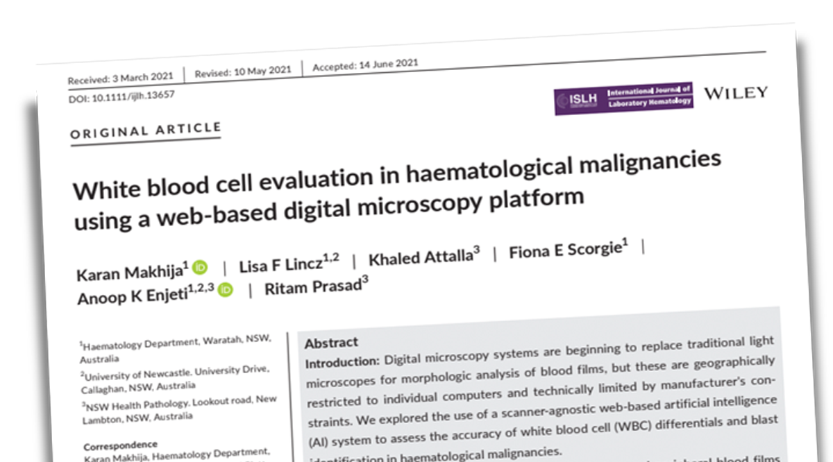
AI-assisted screening tool that reduces technologist read time, speeding triage and determining whether further tests may be needed.
Automated Blood Differential

Overview
Our automated blood differential solution classifies and counts the following objects (for a full list see below):
Challenges & Solutions
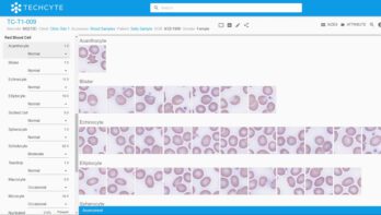
As lab technologists battle eye strain, physical fatigue, distractions, intense workloads and operator biases, their efforts are more prone to human error. AI doesn’t get overworked or tired and delivers consistent results.
Our solution is designed to help technologists quickly eliminate negative blood samples using a more ergonomic computer monitor and allows them to focus more time on positive cases.
How it works
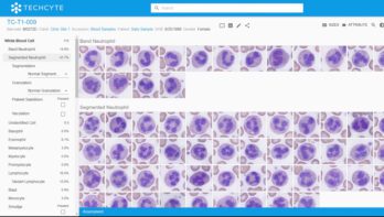
Our Automated blood cell differential solution uses deep machine learning to analyze any good-quality 40x image, identify the monolayer on a normal blood smear, and perform a pre-classification of white blood cells, red blood cells and platelets for a technologist to efficiently review objects of interest prior to signing out the case.
4-step process:
Create slides
Technologists prepare slides following the current CDC guidelines for blood smears, then a standard Romanowsky-type (May Grünwald Giemsa, Wright Giemsa, or Giemsa) staining protocol. Slides are then coverslipped for scanning.
Scan slides
Technicians load slides into the chosen scanner. Slides are then scanned and the resulting images are automatically uploaded to the Techcyte platform for AI analysis. We accept any good-quality 40x image from a compatible scanner (for a list of compatible scanners, see below).
Process images
Our AI algorithm uses a convolutional neural network to identify differentiating features and determine which combinations indicate a certain blood cells or other diagnostically significant objects. It then places them into the most likely classification for technologist review. This algorithm is deterministic, making the same classification every time it is shown the same image. The whole process takes just minutes.
Review results
A technologist logs into Techcyte on any web-enabled device and reviews available samples, confirming presence of objects of interest and, if required, their prevalence. They can also add notes and request a secondary consult. Once complete, results are sent to their LIS.
Cells Identified
- Segmented neutrophils
- Band neutrophils
- Eosinophils
- Basophils
- Lymphocytes
- Monocytes
- Blasts
- Promyelocytes
- Myelocytes
- Metamyelocytes
- Variant lymphocytes
- Smudge cells
- Artifacts
- nRBC
- Unidentified cells
- Segmentation
- Toxic Vacuolation
- Platelet Satellism
- Granulation
- Nucleated red blood cell (nRBC)
- Polychromatophilic RBCs
- Hypochromatophillic RBCs
- Microcytes (user-customizable normal size threshold)
- Macrocytes (user-customizable normal size threshold)
- Target Cells
- Schistocytes (including bite, helmet, horn, triangular, and small fragments)
- Sickle Cells
- Elliptocytes
- Teardrop cells
- Stomatocytes
- Acanthocytes
- Echinocytes
- Spherocytes < 6.5 µm
- Blister Cells
- Platelets
- Large Platelets
- Giant Platelets
- Aggregated Platelets (3 or more platelets grouped together)
Supported scanners

Hamamatsu S360, S20

3DHistech P250, P1000
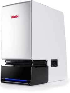
Motic One
Intended Features
- State-of-the-art platform
- AI-proposed images of blood cells and objects of interest, grouped by class and sorted by confidence
- May reduce read times to 30-seconds
- Object counts
- Presence of abnormalities
- Quick drop-down selection of object features
- Case availability after only 7 minutes after scanning.
- No daily cycle of fatigue, distraction, or operator bias
- Levels out sample and stain variations
- Excels at low prevalence samples
- High volume, high reliability scanners produce good quality 40x digital images
Benefits
Partners


