Techcyte’s AI image analysis, coupled with CytoBay’s proprietary stain, results in the first non-invasive, highly accurate detection method which will help make bladder cancer detection and screening more appealing for patients.
Product
Non-Invasive Bladder Cancer Detection

Overview
Our Human Bladder Cancer Detection solution is being developed to be a revolutionary screening tool that helps Pathologists read urine slides in less time and more accurately using an AI-based algorithm trained to detect ICC positive cancer cells.
Our Bladder Cancer solution will classify and count the following object (for a full list see below):
ICC positive low grade cells
ICC positive high grade cells
Uroepithelial Cells
Squamous Cells

Challenges & solutions
Bladder cancer affects over a million people in the US every year. It’s the 4th leading cancer for men. Survivors are supposed to undergo a quarterly biopsy to monitor its progression. This biopsy is very expensive, painful, and invasive for the patient and time-consuming for the Pathologist who analyzes the sample under a microscope.
Our solution is being developed to offer individuals a noninvasive bladder cancer urine test that is pain-free, more accurate, significantly less expensive, allows for quicker results, and is more accessible.
How it works
4-step process:
Create slides
Technicians prepare slides following standard slide preparation protocols and the Cytobay Laboratories chemistry. Slides are then coverslipped for scanning.
Scan slides
Technicians load slides into a compatible whole slide scanner. Slides are then scanned, and the resulting images are automatically uploaded to the Techcyte platform for AI analysis. The Bladder Cancer test accepts any good-quality 40x image from a compatible scanner (for a list of compatible scanners, see below).
Process images
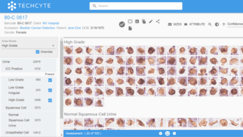
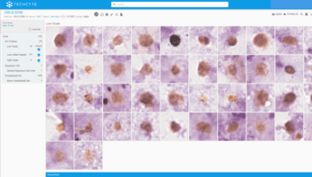
Our AI algorithm uses a convolutional neural network to identify ICC positive low grade, high grade, and other diagnostically significant cells. It then places them into the most likely classification for technologist review. This algorithm is deterministic, making the same classification every time it is shown the same image. The whole process takes just minutes.
Review results
A technologist logs into the Techcyte platform on any web-enabled device and reviews available samples, confirming the presence of objects of interest and, if required, their prevalence. They can also add notes or request an additional consult.
Cells identified
*These claims have not been examined by the FDA.
- High grade urothelial carcinoma
- Low grade urothelial carcinoma
Squamous Cells
Uroepithelial Cells
Supported scanners
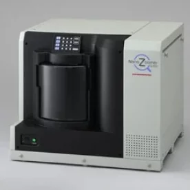
Hamamatsu S360, S20
40x scanning, 20x 0.75 NA objective
3DHistech P250, P1000
40x scanning, 20x 0.75 NA objective
Features
- State-of-the-art platform
- AI-proposed images of ICC positive low and high grade cells, grouped by class and sorted by confidence
- No daily cycle of fatigue, distraction, or confirmation bias
- High volume, high reliability scanners produce 40x equivalent digital images
Benefits
Non-invasive
Pain-free
Inexpensive
Improved accuracy, efficiency
Will help screen for the presence of ICC positive cell
More time available for analyzing positive cases
Reduced technologist stress and fatigue
Improved hiring, training, and retention of lab techs and technologists
Partners
For investigational use only and not for diagnostic use in the US.